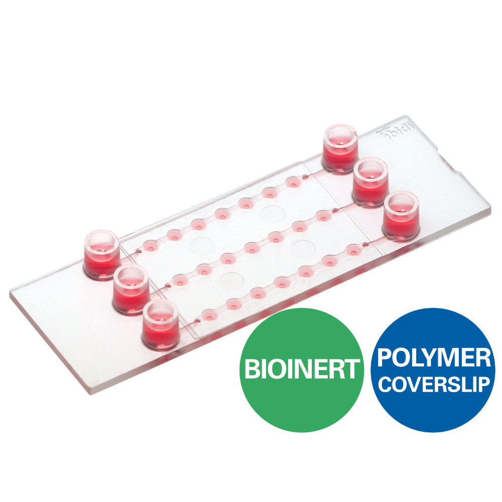
A perfusable channel µ-Slide with 3 x 7 wells for long-term spheroid cultivation and high-end microscopy
- Ideal for the long-term cultivation and perfusion of 3D spheroids or organoids
- Flexible preparation for perfusion: spheroids can be transferred or created directly in the wells
- Imaging chamber with excellent optical quality for high-resolution microscopy
Applications
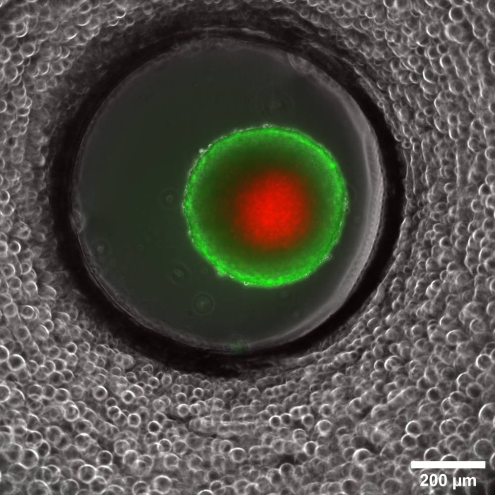
- Cultivation and high-resolution microscopy of three-dimensional cell aggregates (e.g., spheroids and organoids)
- For use with the ibidi Pump System or any other pump device with Luer connectors
- Perfusion of spheroids for fresh medium supply during long-term cultivation and access to spheroid growth kinetics
- Spheroid generation directly in the wells
- Spheroid retrieval for downstream processing (e.g., immunofluorescence, histology, and biochemical assays)
- Perfusion of cell monolayers (adherent cells), suspension cells, or co-cultures
- Organ-on-a-chip setups with up- and downstream metabolization
- Live cell imaging and microscopy of 3D cell aggregates
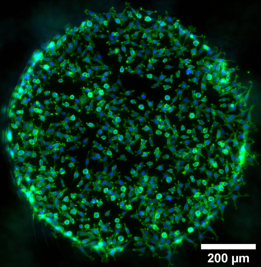
- Immunofluorescence staining and high-resolution fluorescence microscopy of living and fixed cells and cell aggregate
Technical Features
- Optimized well geometry for spheroid/organoid culture and microscopy
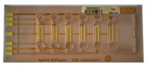
- Analysis of up to 21 samples in one slide
- Perfusion can be applied for optimal nutrient supply during the cultivation of 3D cell aggregates
- Initial open well format allows for easy sample preparation—easily close the wells with a coverslip for the application of flow
- Channels create a simple fluidic connection of the wells
- Luer adapters enable easy pump connection (e.g., to the ibidi Pump System)
- Available with three surfaces:
- Bioinert for complete surface passivation and no cell adhesion
- Uncoated for minimal cell adhesion and suspension cells
- ibiTreat (tissue culture-treated) for optimal cell adhesion
- Sample can be observed through the ibidi Polymer Coverslip bottom using high-resolution fluorescence microscopy
- Compatible with staining and fixation solutions
- Compatible with differential interference contrast (DIC) microscopy when used with a DIC lid
- Made of fully biocompatible materials
