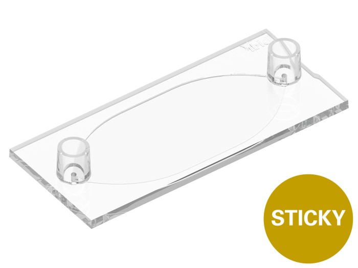
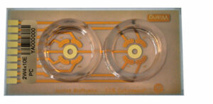
A channel slide designed to support precise immunofluorescence and immunohistochemistry stainings of tissue sections on microscope slides
Applications
- Tissue staining, Immunohistochemistry (IHC), immunocytochemistry (ICC), immunofluorescence (IF) of formalin-fixed, paraffin-embedded (FFPE) tissue
- (Fluorescence) in situ hybridization ((F)ISH)
- DNA-paint on tissue samples
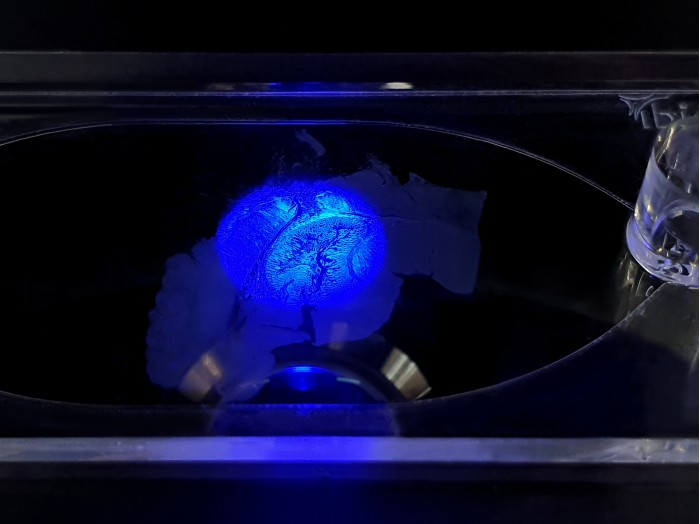
- Microscopy and imaging of slide-mounted tissue
- Parallelization and automation of staining procedures
- Spatial biology research
- Sequential immunostaining
- Multiplex staining
- Other possible applications: fresh frozen sections (cryosectioning) or living tissue
Technical Features
- Creates a channel over slide-mounted tissue to minimize waste of valuable reagents
- Sticky bottom for easy mounting onto glass microscope slide
- Standard Luer ports for easy connection to adapters, tubing, and pumps
- Low volume chamber – saving antibodies and probes
- Low evaporation during staining
- No need for manual hydrophobic barriers by PAP pens
Experimental Workflow
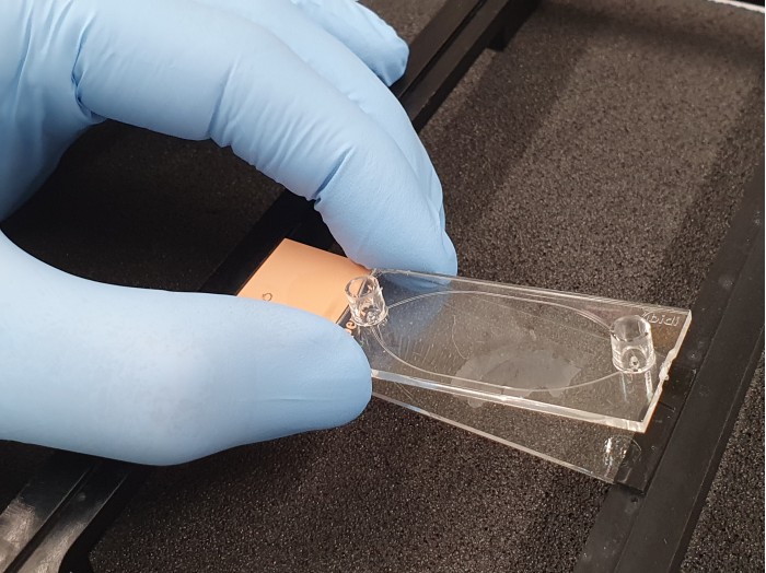
The Sticky-Slide Tissue offers a simple workflow for creating a space-saving channel over a slide-mounted tissue section. After the tissue is deparaffinized and rehydrated, remove the backing and place the sticky slide on the microscope slide to create a convenient staining chamber for the tissue section.
Perform your staining protocol manually with a pipet or multiplex with automated equipment, such as tubing and pumps.
