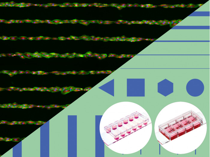
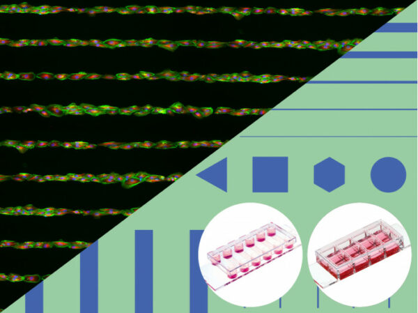
A micropatterned surface with multiple geometries for testing different patterns for the establishment of cell culture assays
Applications
- Determination of a suitable micropatterning geometry for your chosen cell culture application
- Culture and imaging of three-dimensional cell aggregates or cell patch monolayers
- Culture and microscopic analysis of spheroids, organoids, embryoid bodies, and stem cells
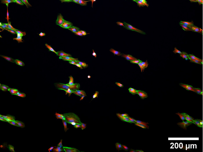
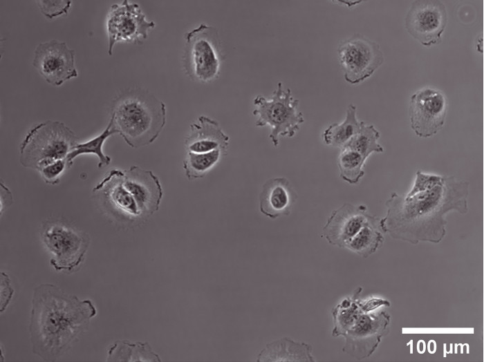
- Analysis of single cells using various approaches (e.g., transfection, proteomics, or metabolic activity tests) with microscopy readout
- Single-cell variability assays (e.g., CAR-T cell activity assay)
- Culture of 3D cell aggregates under perfusion and shear stress (using the µ-Slide VI 0.4 product variation)
- Live cell imaging and fluorescence microscopy
- Immunofluorescence staining and high-resolution fluorescence microscopy of living and fixed cells
Technical Features:
- No cell or protein adhesion
- Long-term stability
- Biologically inert
Specifications
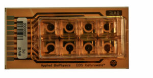
| µ-Slide geometry | See product page µ-Slide 8 Well high or µ-Slide VI 0.4 |
| Binding motif | RGD |
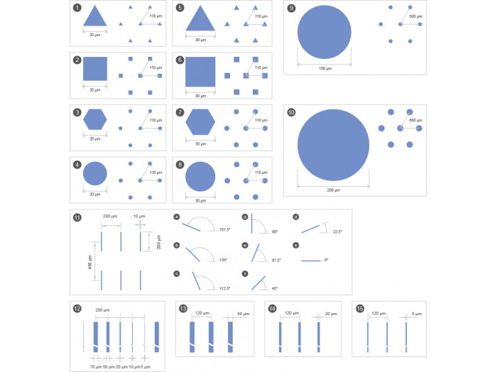
| Pattern shape | 15 variations, see details below |
| Surface passivation | Bioinert |
