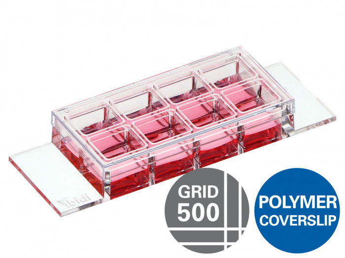
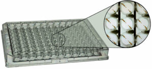
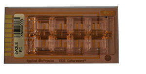
A chambered coverslip with 8 individual wells, high walls, and an imprinted 500 µm cell location grid for use in immunofluorescence and high-end microscopy
- Ideal for locating cells or cell clusters
- Observation of the sample through the polymer coverslip bottom using high resolution microscopy
- Cost-effective experiments using small numbers of cells and low volumes of reagents
- Extra high individual walls to keep cross contamination between wells as low as possible
Applications
- Locating cells or cell clusters, e.g., transfected cells for clone picking
- Counting events per defined area (e.g., for calculating transfection efficiency)
- Providing a reference structure for cell movements
- Cultivation and microscopy of cell cultures
- Transfection assays
- Fluorescence microscopy of living and fixed cells
- Live cell imaging over extended time periods
- Suitable for DIC, when used with a DIC lid
Technical Features
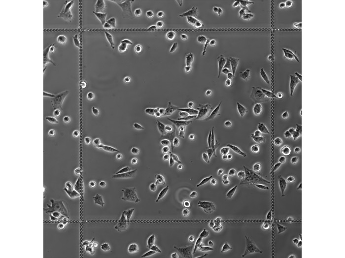
- Grid with a 500 µm repeat distance
- Lettered and numbered fields (A–K; 1–10)
- Open µ-Slide (chambered coverslip) with 8 independent wells
- Individual well walls for minimizing well-to-well crosstalk and contaminations
- Grid clearly visible in phase contrast and bright field microscopy; slightly visible in fluorescence mode
- Excellent optical quality imaging chamber for high-end microscopy
- Compatible with staining and fixation
- ibiTreat surface for optimal cell adhesion
- Biocompatible plastic material—no glue, no leaking
